Standing up for the veterinary profession
08 Aug 2024
25 Aug 2016 | David Gould
Ophthalmology is a discipline that confuses and sometimes intimidates vets in first-opinion practice. In reality, there is no great mystery to clinical ophthalmology.
Ophthalmology is a discipline that confuses and sometimes intimidates vets in first-opinion practice. In reality, there is no great mystery to clinical ophthalmology. True, there is an equipment issue; for conditions such as glaucoma, no matter how much an eye ‘looks’ glaucomatous, without a tonometer you are never going to make an accurate diagnosis, let alone know whether your treatment is effective at lowering the intraocular pressure. But, diagnostic equipment aside, ophthalmology is primarily a discipline of pattern recognition - once you have seen a particular condition you tend to remember it and recognise it the next time a similar case presents to you.
There are, however, a number of mistakes or misunderstandings that seem to crop up fairly regularly in first opinion practice.
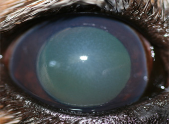
An example of a common misdiagnosis is the failure to identify the presence of corneal oedema. One of the mechanisms for maintaining transparency of the normal cornea is its relative dehydration. This is achieved by a combination of a hydrophobic epithelium which repels ocular surface water, and a sodium/potassium ATPase pump system within the corneal endothelium, which actively removes water from the corneal stroma. A failure in one or other of these systems (either breakdown of the corneal epithelium, such as corneal ulceration, or reduction of the endothelial pump mechanism) leads to corneal oedema.
However, because oedema gives the cornea a cloudy appearance, this condition is too often mistaken for keratitis; a misdiagnosis that can actually be a sight-threatening.
Keratitis is usually associated with ocular surface disease such as corneal ulceration and keratoconjunctivitis sicca. Whilst these are serious conditions in their own right, they are (with the exception of deep ulcers) not usually an emergency requiring urgent diagnosis and treatment if sight is to be saved.
Corneal oedema, on the other hand, can be a sign of severe intraocular disease that, if missed, can lead to permanent blindness – examples including glaucoma, lens luxation, or anterior uveitis.
But whilst corneal oedema and keratitis can look similar at first glance, they are actually quite different in appearance. Corneal oedema gives a characteristic ‘stippled’ appearance (which often reminds me of the stippling on the surface of an orange).


This stippling is due to hydration of the stromal collagen fibrils, which then swell and separate, scattering light to cause this characteristic appearance.
Compare this to non-ulcerative keratitis (Figure 3) which also causes corneal clouding, but without the stippled appearance, and often with additional signs such as vascularisation or pigmentation.

So if you see the tell-tale stippled appearance of corneal oedema, first perform fluorescein-staining to rule out corneal ulceration, and then look for an intraocular cause, with particular emphasis of identifying potentially serious and blinding causes such as glaucoma, lens luxation or anterior uveitis. Thus pattern recognition, in this case the ability to recognise the typical appearance of corneal oedema, is vital if these conditions are to be recognised and treated in time.
I will be addressing the most common ophthalmology mistakes and misunderstandings on BVA’s ophthalmology course on 4 October. Questions we will be addressing include:
Please note this course is now fully booked. If you would like to be added to the waiting list please email [email protected].
Get tailored news in your inbox and online, plus access to our journals, resources and support services, join the BVA.
Join Us Today